Oleh: Thalia Kaylyn Averil
Tomografi komputer jantung, atau Cardiac CT, merupakan teknologi pencitraan medis yang menghasilkan gambar tiga dimensi (3D) berkualitas tinggi dari jantung, pembuluh darah, dan struktur terkait. Prosedur ini menggunakan sinar X pada berbagai sudut untuk menciptakan gambaran rinci. Selain itu, Cardiac CT dapat digunakan untuk mengevaluasi berbagai komponen, seperti ruang jantung, otot jantung, katup, perikardium, arteri koroner, serta arteri dan vena besar.
Fungsi utamanya bertujuan untuk melihat lebih jelas struktur jantung dan pembuluh darah terkait. Alat diagnostik ini dapat digunakan untuk menentukan penyebab sesak napas dan nyeri dada, mengamati jantung sebelum dan setelah dilakukan prosedur medis, memeriksa kelainan yang terjadi di arteri koroner dan arteri-arteri besar, mengamati massa atau tumor yang berada di jantung atau sekitarnya, memeriksa adanya cairan atau pengapuran di sekitar lapisan yang melindungi jantung, mengevaluasi kinerja katup jantung, atau memeriksa penyakit jantung bawaan.
Untuk proses pengambilan gambar sendiri dapat dilakukan dengan atau tanpa menggunakan kontras; Cardiac CT yang dilakukan dengan kontras disebut juga dengan coronary CT Angiography (cCTA). Coronary CT Angiography memberikan informasi mengenai anatomi jantung dan pembuluh darah jantung, terutama untuk evaluasi penyakit arteri koroner dan penyakit kardiovaskular lainnya.
Namun, ada beberapa keterbatasan yang dimiliki oleh Traditional Cardiac CT, yaitu dalam kecepatan perolehan gambar, resolusi spasial, dan resolusi jaringan lunak. Saat ini, CT-Scan tradisional yang ada masih belum optimal dalam mencitrakan arteri koroner dan stent yang telah mengalami pengapuran berat. Selain itu, lemak dan plak nonkalsifikasi di sekitar arteri koroner masih sulit dibedakan karena terbatasnya resolusi spasial pada Cardiac CT. cCTA juga memiliki beberapa kekurangan, yaitu kurang efektif untuk arteri kecil (kurang dari 2 mm), kontras tinggi (seperti kalsifikasi atau stent), dan kontras rendah (seperti plak nonkalsifikasi).
Oleh karena itu, teknologi terbaru bernama photon-counting CT (PCCT) memiliki resolusi spasial yang lebih baik sehingga dapat menangkap gambaran jantung yang lebih jelas. Dengan meningkatkan resolusi spasial, PCCT dapat mengukur sumbatan dan menilai timbunan lemak di pembuluh darah secara lebih akurat dari Cardiac CT. Selain itu, masalah-masalah seperti gambaran buram di sekitar timbunan kalsium dan gambaran yang kurang baik dalam menampilkan jaringan lunak dapat diatasi dengan PCCT.
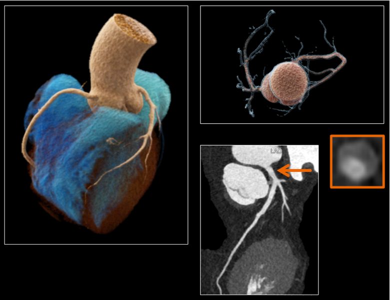
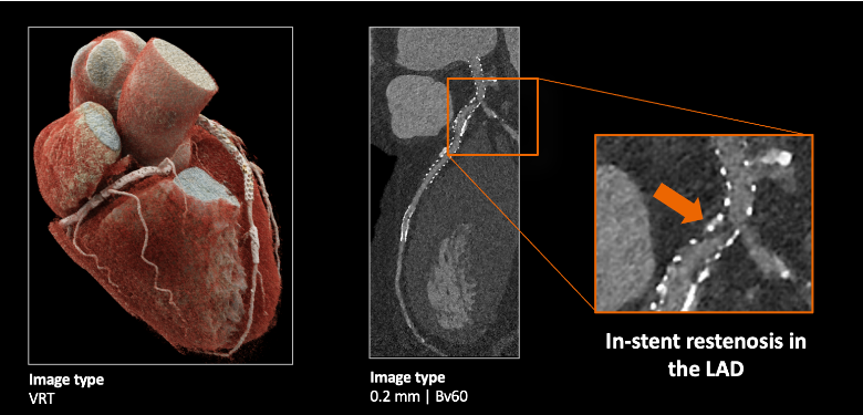
Gambar 1. Hasil dari photon-counting CT.
Lalu, bagaimana cara kerja dari Photon-Counting CT (PCCT)?
Photon-counting CT dan Traditional Cardiac CT berbeda dalam pemrosesan foton yang berasal dari sinar-X. PCCT menggunakan detektor yang dapat menghitung masing-masing foton dan menentukan tingkat energinya, berbeda dengan cardiac CT yang menggunakan detektor pengintegrasi energi. Detektor yang digunakan oleh PCCT menghitung dan mengklasifikasikan foton berdasarkan energinya saat sinar-X melewati tubuh sehingga dapat memberikan informasi akurat tentang komposisi jaringan. Traditional Cardiac CT bekerja dengan mengubah sinar-X menjadi cahaya dan kemudian mengubah cahaya menjadi sinyal elektronik, tetapi PCCT dapat mengurangi konversi cahaya sehingga meningkatkan efisiensi sinyal dan mengurangi dosis radiasi yang digunakan selama prosedur PCCT.
Keunggulan Photon-Counting CT dibandingkan dengan Traditional Cardiac CT adalah:
- Resolusi Spasial yang Lebih Tinggi: Menawarkan peningkatan resolusi spasial, sehingga gambaran anatomi jantung dan pembuluh darah sekitarnya dapat divisualisasikan dengan lebih baik.
- Dosis Radiasi yang Lebih Rendah: Memungkinkan pelaksanaan pemindaian dengan dosis radiasi yang signifikan lebih rendah daripada Traditional Cardiac CT, sementara tetap menghasilkan detail gambar yang tinggi.
- Lebih nyaman untuk Pasien: Menghasilkan lebih sedikit kebisingan dibandingkan Traditional Cardiac CT, menciptakan pengalaman yang lebih nyaman bagi pasien.
Berikut adalah perbandingan antara cardiac CT dengan photon-counting CT.
| Traditional CT | Photon-Counting CT |
| Memiliki resoulsi spasial yang terbatas sehingga kurang direkomendasikan untuk mencitrakan struktur-struktur anatomi yang kecil. | Memiliki resolusi spasial yang lebih baik sehingga gambaran yang dihasilkan lebih jelas dan detail, terutama untuk struktur-struktur seperti pembuluh darah yang kecil. |
| Membutuhkan dosis radiasi yang lebih tinggi untuk mencapai kualitas gambar yang diinginkan. | Membutuhkan dosis radiasi yang lebih kecil daripada traditional CT karena dapat mengurangi konversi cahaya. |
| Bekerja dengan menggunakan detektor pengintegrasi energi foton dari sinar-X. | Bekerja dengan menggunakan detektor yang dapat menghitung dan menentukan tingkat energi masing-masing foton dari sinar-X sehingga lebih akurat dalam menentukan komposisi jaringan. |
| Memproduksi bising yang lebih banyak selama prosedur. | Memproduksi bising yang lebih sedikit selama prosedur. |
Sebagai institusi yang selalu berkomitmen dalam menyediakan layanan kesehatan terdepan, Rumah Sakit Abdi Waluyo dengan bangga menghadirkan Photon-Counting CT sebagai salah satu pelayanan unggulannya. Kami percaya, dengan teknologi ini, pasien akan mendapatkan diagnosa yang lebih akurat dan pengalaman perawatan yang lebih baik.
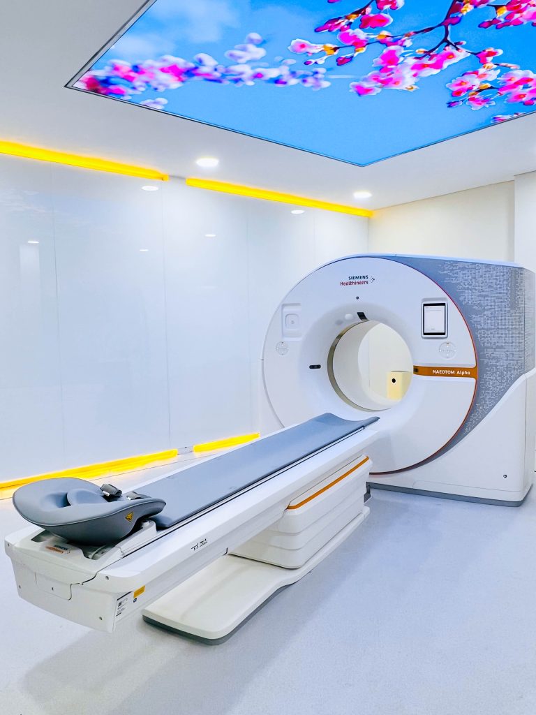
Gambar 2. Photon-counting CT di Rumah Sakit Abdi Waluyo.
Referensi:
- Cleveland Clinic. Cardiac computed tomography (CT) scan [Internet]. Cleveland: Cleveland Clinic; date of publication unknown [reviewed 2022 Feb 28] [cited 2024 Jan 9]. Available from: https://my.clevelandclinic.org/health/diagnostics/16834-cardiac-computed-tomography
- Si-Mohamed SA, Boccalini S, Lacombe H, Diaw A, Varasteh M, Rodesch P, et al. Coronary CT angiography with photon-counting CT: first-in-human results. RSNA [Internet]. 2022 Feb 15 [cited 2024 Jan 9];303(2). Available from: https://pubs.rsna.org/doi/full/10.1148/radiol.211780
- Leng S, Bruesewitz M, Tao S, Rajendran K, Halaweish AF, Campeau NG, et al. Photon-counting detector CT: system design and clinical applications of an emerging technology. RSNA [Internet]. 2019 May 6 [cited 2024 Jan 9];39(3). Available from: https://pubs.rsna.org/doi/full/10.1148/rg.2019180115
- Meloni A, Cademartiri F, Positano V, Celi S, Berti S, Clemente A, La Grutta L, Saba L, Bossone E, Cavaliere C, Punzo B, Maffei E. Cardiovascular Applications of Photon-Counting CT Technology: A Revolutionary New Diagnostic Step. J Cardiovasc Dev Dis. 2023 Aug 25;10(9):363. Available from: https://www.ncbi.nlm.nih.gov/pmc/articles/PMC10531582/